SOPs

Validation of Laminar Air Flow and Its SOP

Validation of Laminar Air Flow; Purpose

To validate Laminar Air Flow in order to support of processing area and Microbiology Test.

Validation of Laminar Air Flow; Scope

This SOP applies for validation of Laminar Air Flow used in Processing area and Microbiology Section XX Pharmaceuticals Ltd.

Definitions/Abbreviation :

Validation: Validation is the established documented evidence, which provides a high degree of assurance that a specific process will consistently produce a product meeting its predetermined specifications & quality attributes.

DOP Test: Dioctyl Pthalate is a combustible non-toxic colorless oily liquid with slight odor. This chemical is used in HEPA filter integrity test at vaporized condition by DOP Test Meter.

[] HEPA : High Efficiency Particulate Air

[] CSDA : Casein Soyabean Digest Agar

[] SDA    : Sabouraud Dextrose Agar

[] LAF    : Laminar Air Flow

 Responsibilities

The roles and responsibility is as follows

Executive/ Senior Executive, Microbiology

Perform Microbiology Air monitoring of Laminar Air Flow & report preparation.

Executive/ Senior Executive, Engineering

Perform Air Velocity Test, DOP test and report preparation.

Microbiology/ Assistant Manager, Engineering

Ensure Laminar Air Flow Validation, documentation and application of sound technical information.

Head of Quality Assurance

Take initiative to Approval of SOP

Procedure:

Instructions
  • Wear protective items such as sterile latex free gloves, laboratory coat and eye protection (if required).
  • Disinfect whole surface of all apparatus with the help of 70% IPA or any others disinfectants before transferring into Laminar Air Flow.
  • Make sure that all personal ornaments, cell phone are left to prevent unauthorized contamination, before working under Laminar Air Flow.
Microbiological Monitoring
  • Start Laminar Air flow at least before 30 minutes of validation.
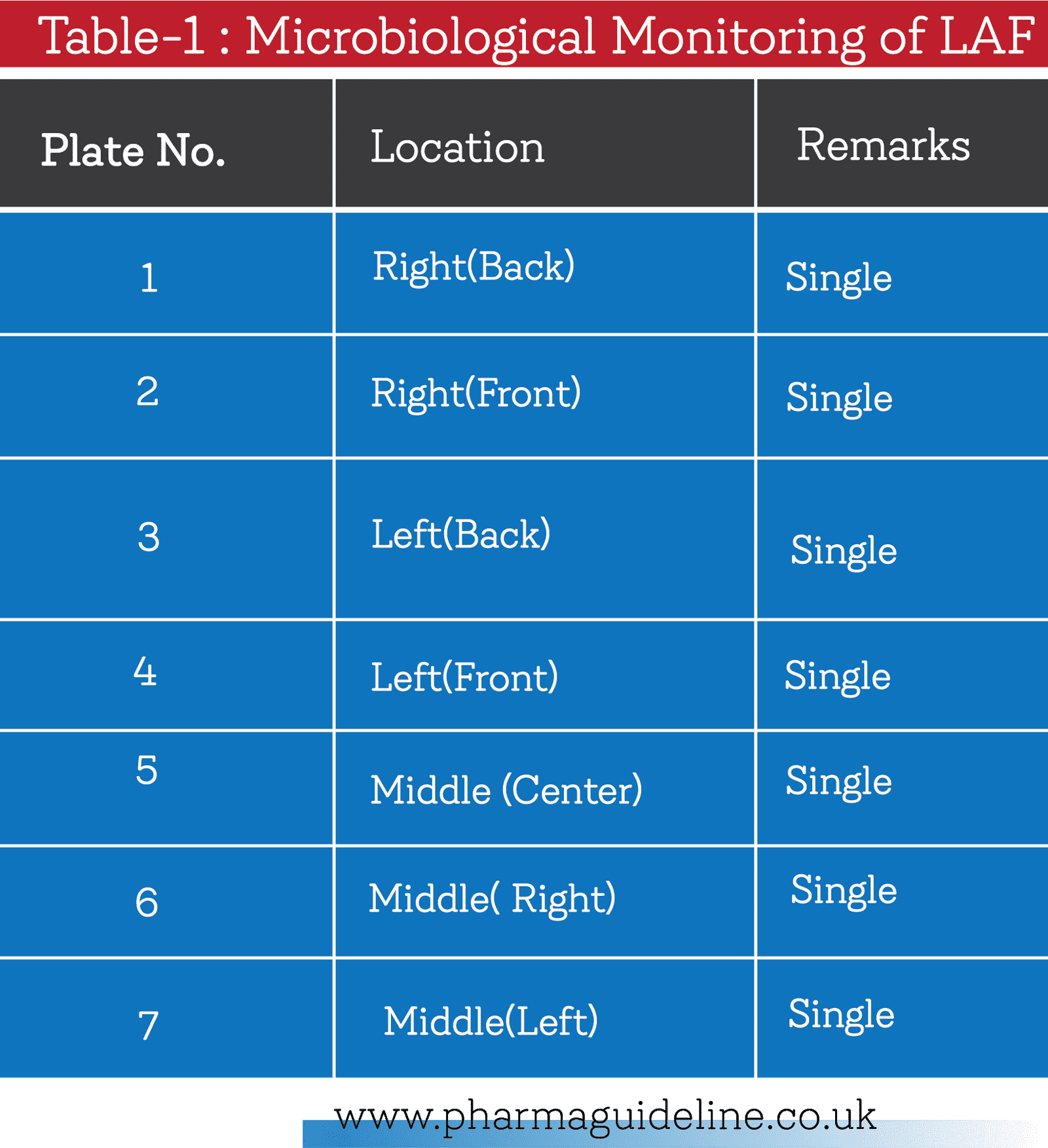
  • Select sampling point as mentioned on Table-1.
  • Expose sterile CSDA and SDA plate for 30 minutes under LAFWS at different location as per sampling points.
  • After sampling, cover all plates with the glass lids.
  • Incubate all CSDA plate at (30 to 35)0C for 72 hours and SDA plate at (22 to 25)0C for 5 days including positive control & negative control plate.
  • After incubation, count the CFU per plate.
  • Report in Laminar Air Flow Validation Report, Annexure-I.
  • Perform this test yearly.
 Detection of Air Velocity
  • Disinfect outer surface of Anemometer with the help of 70% IPA before transferring into LAF.
  • Measure distance 6” from HEPA.
  • Take air flow reading of HEPA of LAF by moving smoker from left to right & top to down.
  • Check that any deviation of reading found for air velocity. If found mark that point.
  • Report Air Velocity & Filter Integrity Test Report of Laminar Air Flow, Annexure-II.

Filter Integrity Test

  • Set DOP Test Meter with DOP test port of LAF.
  • Run machine as per SOP of DOP Test Meter.
  • Take air by smoker of DOP Test & record the reading.
  • Check any deviation of reading.
  • Report Air Velocity & Filter Integrity Test Report of Laminar Air Flow, Annexure-II.
Validation Frequency

Perform LAF Validation once in a year.

Validation of Laminar Air Flow


Corrective Action:

[] If growth obvers in anyone plate, inform the test result to concerned department & Engineering Department for corrective action.

[] If the test result found out of specification in Air Velocity Test or Filter Integrity Test, Engineering department shall take necessary action.

[] Microbiology Section and Engineering Department shall perform the complete test.

Download All Annexure Here:

Annexure I Air Velocity & Filter Integrity Test Report of Laminar Air Flow Biosafety Cabinet
Annexure II Laminar Air Flow Bio-safety Cabinet Validation Report

Validation of Laminar Air Flow and Its SOP Read More »

Microbial Examination of Empty Bottle, Cap & Stopper

Microbial Examination of Empty Bottle, Cap & Stopper; Purpose

To confirm that the bacterial & fungal count & specified microorganisms into empty bottle during filling are within In-house specification.

Scope

This SOP is applicable for microbiological test of empty bottle during filling in Microbiology Section at XX Pharmaceuticals Ltd.

Definitions/Abbreviation

[] CSDA: Casein Soyabean Digest Agar

[] CSDM: Casein Soyabean Digest Medium

[] SDA  :  Sabouraud Dextrose Agar

[] TAMC: Total Aerobic Microbial Count

[] TYMC: Total Yeast & Mould Count

Responsibilities

The roles and responsibility is as follows

Executive, Microbiology

Sample collection, analysis and documentation.

Manager, Microbiology/Quality Control

Ensure analysis of empty bottles, documentation and application of sound technical information.

Head of Quality Assurance

Take initiative to Approval of this SOP

Procedure

Instructions
  • When enter into the test area, wear sterile latex free gloves, mask, laboratory coat & eye protection (if required).
  • Make sure that all personal ornaments cell phone are left to prevent unauthorized contamination, before entrance into test area.
  • Move always gently and never move vigorously into the test area.

General Requirements for the test

Glass Apparatus
  • Sterilized 90 mm Glass Petridish
  • Screw capped Conical Flask 100 ml
  • Screw Capped Test Tube
  • Volumetric Flask 1000 ml
  • Volumetric Flask 500 ml
  • Pipette 2 ml, 10 ml
Media and Reagents
  • Casein Soyabean Digest Agar(CSDA)
  • Casein Soyabean Digest Medium(CSDM)
  • Mac-Conkey Broth
  • MacConkey Agar
  • Neutralized Peptone
  • Sabouraud Dextrose Agar
Others Requirements
  • Surgical Gloves
  • Surgical Cotton
  • 70% IPA or ethanol

Enumeration Method(TAMC & TYMC)

This test quantifies the enumeration of mesophillic bacteria & fungi that may grow under aerobic conditions.

Test Conditions
  • Disinfectant both hands, bottles surface, Laminar Air Flow workstation with 70% IPA or 70% ethanol before starting test.
  • Carry out the test under Laminar Air Flow to avoid contamination.
Culture Media Preparation
  • Prepare different culture media as per requirement.
  • Weigh accurate amount mentioned in the manufacturer label into appropriate flask.
  • Bring to boil completely to dissolve media.
  • Sterilize at 1210C for 15 minutes or as per manufacturer label.
  • Store prepared culture media in air tight flask at 2 to 80
Stock Buffer Solution
  • Take 34 g of Potassium Dihydrogen Phosphate[KH2PO4] in a 1000 ml volumetric flask.
  • Dissolve in 500 ml of Purified Water, adjust to pH 7.2 ± 0.2 & dilute to 1000ml with Purified Water.
  • Dispense 90 ml into each screw capped flask.
  • Sterilize at 1210C for 15 minutes.
  • Store prepared buffer at 2 to 80C for a validated period.
Glassware Cleaning & Sterilization
  • Clean all glassware by 1% detergent initially & then rinse with sufficient tap water.
  • Rinse finally with sufficient Purified Water to remove residual content of detergent.
  • Sterilize all glassware at 2000C for 1 hour.
Sample Size
  • Collect 5 empty sealed bottles from each batch during filling of the products at three stages of Starting, Middle and Ending of operation.

Test Method

  • Carry out anyone from the following mentioned method
Pour plate method
  • Transfer all bottles under Laminar Air Flow.
  • Deseal the cap of all bottles.
  • Add 10 ml sterile meat peptone or phosphate buffer pH 7.0 ± 0.2.
  • Cap the bottles & shake well to mix properly.
  • Mark all petridish as product name, batch number, plate name (CSDA/ SDA) and bottle number & test date.
  • Take 1 ml of rinsing solution from each bottle & pour into two 90 mm sterilized petridish.
  • Add 15-20 ml CSDA into one plate & add 1 ml SDA into another plate.
  • Maintain same manner for the rest 4 bottles.
  • Mix sample with the media by tilting & rotating the plate.
  • Allow to solidify all plates & invert after solidification.
  • Incubate CSDA at 30 to 350C for 3 to 5 days & at 20 to 250C for 5 to 7 days.
  • After incubation, calculate number of cfu per bottle.
Negative Control

Use the diluents as sample in place of test preparations and follow the steps mentioned steps.

Membrane Filtration Method
  • Prepare sample as per Pour Plate Method
  • Filter whole rinsing solution of all bottles individually through 0.45 µm and transfer the filter paper to the surface of CSDA slant for bacterial count and SDA slant for yeast & mold count.
  • Invert plates & incubate all CSDA plates at 30 to 350C for 3 to 5 days & SDA plates at 20 to 250C for 5 to 7 days.
  • After incubation, count the colony of each plate.
  • Calculate number of cfu per bottle.
Negative Control

Use the diluents as sample in place of test preparations and follow the steps mentioned steps.

Surface spread Method
  • Spread not less than 0.1 ml of rinsing solution on surface of two CSDA & two SDA Plate.
  • Dry all plates under Laminar Air Flow work station.
  • Incubate CSDA at (30 to 35)0C for 3 to 5 days & at (20 to 25)0C for (5 to 7) days.
  • After incubation, calculate number of cfu per bottle.
Negative Control

Use the diluents as sample in place of test preparations and follow the steps mentioned steps.

Interpretation of the results
  • The bottles are passed if the observed count is less than specified count.
  • The product is failed if the observed count is greater than specified count of that product.
Test Control
  • Negative control must be negative growth. If found growth in negative control, the test is invalid.

Test for Specified Microorganisms

Suitability of Test Method
  • Add each test strain separately not more than 100 cfu at the time of bottle test mixing with culture media as per standard operating procedure of Suitability of Microbial Count Method.
  • The test is suitable if growth found the specific microorganisms. The test is not suitable if no growth found the specific microorganisms. In that case, add any neutralizer or increase dilution for removal any inhibition of product.
 Test for E. coli
  • Add 10 ml of test sample to 90 ml of CSDM. Incubate at (30 to 35)0C for 18 to 24 hours.
  • Shake the container and transfer 1 ml of CSDM to 100 ml of MacConkey Broth.
  • Incubate at (42 to 44)0C for 24 hours.
  • Sub culture on MacConkey Agar plate from MacConkey broth.
  • Incubate at (30 to 35)0C for (18 to 72) hours.
  • The product complies with the test for E. coli if no red colonies are present with precipitated zone & the biochemical tests are negative.
Test Report Preparation

Microbial Examination Report of  Cap & Stopper, Annexure-I.

Microbial Examination Report of Empty Bottle, Annexure-II.

Download All Annexure Here

 Microbial Examination Report of Cap & Stopper Annexure-I.
 Microbial Examination Report of Empty Bottle Annexure-II.

Microbial Examination of Empty Bottle, Cap & Stopper Read More »

Microbiology Analysis of Surface Swab and its SOP

Microbiology Analysis of Surface Swab; Purpose:

To confirm that the processing area & processed equipment’s are free from any intolerable microorganisms & total aerobic count are within specification.

 Microbiology Analysis of Surface Swab; Scope:

This SOP applies to detect or count of bacteria and Yeast/Mold in Non-sterile Processing, Sterile Processing Area & Testing area of Microbiology laboratory at XX Pharmaceuticals Ltd.

 Definition:  

Swab test & Finger Printing are to check with a view to take timely corrective measures for maintaining a favorable manufacturing environment, minimizing the risk of contamination of the products.

Responsibilities:

The roles and responsibility is as follows

Executive/ Senior Executive, Microbiology

Sample collection, analysis of swab and document preparation

Manager, Microbiology

Confirm sampling, analysis, documentation & application of updated technical information.

Head of Quality Assurance

Take initiative to Approval of this SOP

Procedure:

Instructions
  • Disinfect with the help of 70% IPA/Ethanol the whole outer surface of machine & all apparatus.
  • Wear gloves, mask & sterilized garments before entrance into aseptic area.
  • Minimize the movement into aseptic area or others sampling area.
Swab Test of Garments/Floor/Wall/Equipment
Sterilization of Swab Kits and Culture Media
  • Prepare swab kits with cotton bud as per requirement.
  • Roll tightly a small amount of cotton of surgical grade at one end of the glass rod.
  • Place cotton bud in a narrow test tube.
  • Plug end of test tube with the help of non-absorbent cotton.
  • Prepare CSDA [Casein Soybean Digest Agar], CSDB [Casein Soybean Digest Broth] according to the requirement of the test. Distribute 50ml of CSDB in each conical flask.
  • Sterilize media & test tubes containing cotton bud & Template steel plate at 1210C & 15lbs pressure for 15 minutes in an autoclave.
Swab Collection
  • Select a steel template of 5 x 5 cm. size.
  • Sterilize steel template by alcohol flaming before the test
  • Wipe cotton bud slowly & firmly in interior direction of steel template on the surface, selected for test.
  • Rotate cotton bud against direction of the overall wiping movement.
  • Repeat process for three times.
Collected Sample Preservation
  • Perform test within 30 minutes after collection of sample.
  • Preserve sample at 2 to 80C at not more than 12 hours if test is not performed within 30 minutes.
 Test Method
  • Place swab immediately in a bottle containing 50ml of CSDB.
  • Pull cotton free in the medium.
  • Shake bottle containing swab for sometime in a shaker.
  • Pour 1ml of diluent into sterile petridishes with aid of a sterile pipette.
  • Add 20 to 25ml of Tryptone Soy Agar at about 45 to 500C to the plate.
  • Mix medium with sample by rotating petridish.
  • Allow medium to solidify & then keep plate for incubation at 370C for 48 hours.
  • After incubation period count number of colonies either by colony counter or visual inspection.
  • Carry out the test in every week.
Interpretation of Test Result
  • Report swab test if test result found in Swab Test Report, Annexure-I.
  • If test result found out of specification, repeat the test.
  • Recollect swab sample from same area or same person.
  • Carry whole test as previously performed
  • If test result found within specification as per table-1, report the test result in Annexure-I.
Personnel Finger Printing :
Culture Media Preparation and Sterilization
  • Prepare CSDA [Casein Soybean Digest Agar] for the test according to the requirement of the test following the instructions of the respective manufacturer.
  • Autoclave medium at 1210C & 15lbs pressure for 15 minutes.
  • Pour approximately (20 to 25) ml of medium in each plate after autoclaving.
  • Allow medium to solidify & then keep in refrigerator until use.
  • Dry surface of agar media.
  • Do not use prepared plate after 72 hours.
  • Take medium aseptically to respective department.
Collection of Finger Print
  • Take finger print of both the hands, of each person working in the aseptic area in the separate plate.
  • Mark each plate with person’s name, area /room no., hand (right/left) and date.
  • Aseptically bring plates back to microbiology laboratory
  • Incubate plates at (30 to 35)0C for 48 hours.
  • After incubation count number of colony forming unit formed on each plate either by visual examination or by colony counter.
  • Carry out test every week for sterile process operator & once in a month for Non-sterile process operator.
 Interpretation of Test Result
  • Report swab test if test result found in Swab Test Report, Annexure-I.
  • If test result found out of specification, repeat the test.
  • Recollect swab sample from same area or same person.
  • Carry whole test as previously performed
  • If test result found within specification as per table-1, report the test result in Annexure-I.
Out of Specification

If test result found out of specification, send test report to concerned Sectional Head for corrective action.

After corrective action, repeat the test for specific area or person.

Download the All Annexure Here

Annexure 1 Swab (Microbial) Test Report
Annexure II Finger Print Test Report

Microbiology Analysis of Surface Swab and its SOP Read More »

Maintain and preserve of standard culture and its SOP

Maintain and preserve of standard culture; Purpose

To maintain & preserve the standard culture in order to use in specific microbiology test.

Maintain and preserve of standard culture;Scope

This SOP applies to preserve & maintain stock standard culture in Microbiology Laboratory of at XX Pharmaceuticals Ltd.

Definitions

Standard Culture

Standard culture is a specific microorganism of a specific strain that is recognized by BP/USP/Eur. Ph.  and certified by  ATCC/NCTC or any other standard culture bank. That culture is used in the different microbiological test such as Growth Promotion Test, Biological Assay of Antibiotics, Sterilizer Validation, Antimicrobial Preservative Effectiveness Test, Microbial Count suitability Test & Sterility Test Validation.

  • ATCC: American Type Culture Collection
  • NCTC: National Type Culture Collection
Responsibilities
Executive/ Senior Executive, Microbiology
  • Preparation of Culture Media, sub-culture, preservation and record keeping.
  • Follow the instructions of this procedure correctly.
Manager, Microbiology
  • Ensure that this procedure is kept up to date.
  • Ensure culture maintenance activities & proper documentation
  • Ensure appropriate personnel from the section are trained on this procedure.
  • Confirm that SOP is technically sound & reflects the required working practices.
Head of Quality Assurance

Take initiative to approval of this SOP

Procedure:

Instructions
  • Standard microorganisms are usually non-pathogenic but those are unscrupulous pathogen. So before handling it, wear protective items such as sterile latex free gloves, laboratory coat and eye protection (if necessary).
  • Move always gently. Don’t move vigorously into the test area.
  • Disinfect the whole surface with the help of 70% IPA or any others suitable disinfectants. After completion of sub-culture or transfer of pellets.
  • Take away all used items those are directly contacted with standard micro-organisms and Leave it at designated containers safely.
  • Confirm the waste container is tightly capped until autoclaving.
  • Never touch the apparatus directly in open hands those are used.
  • Make sure that all personal ornaments, cell phone are left before entrance into the test room, to prevent unwanted contamination.
Sterilization of Apparatus & Glassware:
  • Sterilize all glassware plus Latin Square Plate and others heat stable apparatus at 2000C for 60 minutes in hot air oven using a validated process.
  • Use the glassware when the temperature reduce to 400
Preparation of Culture Media:
  • Select media & diluents as per instruction of BP or USP.
  • Prepare required amount of Culture media of CSDA [Casein Soybean Digest Agar] & CSDB [Casein Soybean Digest Broth] and SDA [Sabouraud Dextrose Agar] as per indication by manufacturer instructions.
  • Bring to boil to dissolve it completely.
  • Distribute 10 ml of the media into each screw capped test tube.
  • Sterilize at 1210C for 15 minutes.
  • Cool media approximately to (45 to 50)0
  • Allow to solidify agar media to prepare slant.
  • Transfer broth media flasks into the test room.
  • Dry surface of agar slant keeping into Laminar Air flow or Bio-safety Cabinet.
  • Incubate sterilized CSDA [Casein Soyabean Digest Agar]/broth at (30 to 35)0C for 48 hours and SDA [Sabauroud Dextrose Agar] for 5 days at (22 to 25)0C for checking sterility of the media.
  • After incubation observe each test tube for growth. If no growth found, the media is suitable for use.
  • Store media at (2 to 8)0C into refrigerator until it use.
Preparation of Standard Microorganism:
Test Conditions

[] Disinfectant the surface of all equipments including LAF or BSC.
[] Rub hands with the help of 70% IPA.
[] Always maintain aseptic condition during handling of standard microorganisms.

Inoculation Technique
  • Opening of Standard Culture vial/ampoule
  • Read carefully label’s instructions of standard culture vial/ampoule.
  • Aseptically break the ampoule/vial under Laminar Air Flow.
  • Remove pellets from vial/ampoule aseptically as per instruction of the label.
  • Tight the container for the next time use after removing pellets.
Inoculation of Standard Microorganisms:
  • Label on the each tube with the name of standard culture.
  • Reconstitute standard culture pellets as per instructions of manufacturer.
  • Aseptically Transfer 1 or 2 pellets of dehydrated culture of bacteria and fungus in the test tubes containing CSB.
  • Incubate test tubes containing bacterial culture at (30 to 35)0C for 3 days and the tubes containing fungi culture at (20 to 25)0C for 5 days. This will serve as mother culture.
  • After incubation, observe the growth of standard culture that should be turbid or settle growth.
  • Transfer & streak from the broth culture on surface of agar slant.
  • Incubate test tubes containing CSA at (30 to 35)0C for 3 days & the SDA tubes containing fungi culture at 20 to 250C for 5 days.
  • Observe good growth, then preserve it at (2 to 8)0C in refrigerator with proper labeling.
  • Discard previous culture by autoclaving at 1210C for 30 minutes.
  • Subculture microorganism to freshly prepared culture media once in a month.
Preparation of spore suspension:
  • Transfer sterilized media under Laminar Air Flow or Bio-safety Cabinet.
  • Keep plate for incubation at 350C for 7 days.
  • After incubation period take out culture with aid of sterilized glass beads & pre-sterilized diluents (0.001 g/L containing Manganese sulfate) by rotating plate.
  • Pour culture suspension along with few of those glass beads in a 100ml flask containing 50ml of sterilized diluents.
  • Heat culture suspension at 700C for 30 minutes or 800C for 10 minutes in a water bath.
  • Cool suspension & then keep inside a refrigerator not exceeding at 40C
  • Don’t use spore suspension more than 60 days.
Preparation of standard culture suspension (Vegetative form):
  • Prepare culture suspension by regular subculture on Nutrient Agar slant or Sabouraud Dextrose Agar.
  • Before using a culture maintained in a slant, subculture the organism in another slant containing a specified medium.
  • Keep the slant for incubation at (30 to 35)0C for bacteria for (24 to 30) hours and at (20 to 25)0C for fungi for (3 to 5) days.
  • Store slant in the refrigerator not exceeding at 40C if not used immediately.
  • Don’t use this suspension at more than 7 days.
Stock Maintenance of Standard Microorganisms:
  • Check expiry date before use of standard microorganisms.
  • Don’t use if the expiry date is exceeded & discard it after autoclaving.
  • Raise requisition for standard culture before exceed of expiry date.
 Report Preparation

Maintain Register of standard culture maintenance Record, Annexure-I.

Download Annexure:

Download the annexure here: Standard Culture Maintenance Record

Maintain and preserve of standard culture and its SOP Read More »

Identification of Microorganisms and its SOP

Identification of Microorganisms; Purpose

To identify Microorganisms.

Identification of Microorganisms; Scope

This SOP applies for identification of Microorganisms in Microbiology Section at XX Pharmaceuticals Ltd.

Definitions
GN-ID System

The GN-ID system employs 12(GNA) or 24(GN A+B) standardized biochemical substrates in microwells to identify the family of Enterobacteriaceae & other non-fastidious Gram negative bacilli (Oxidase negative and positive). The kit is planned for professional laboratory use only.

[] CPG: Colony Pigmentation.
[] CAT : Coagulate test
[] LAT : Latex Agglutination Test
[] ONPG : Ortho-Nitrophenyl β-Galactoside
[] PYR : PYRROLIDONYL ARYLAMIDASE
[] TDA : Tryptophan Deaminase Agent( Indolepyruvic Acid)
[] VP : Voges-Proskauer

Identification of Microorganisms;Responsibilities:
Executive/ Sr. Executive, Microbiology

Selection of culture and identification of Microorganisms.

Assistant Manager, Microbiology

Ensure identification and application of sound technical information.

Head of Quality Assurance

Take initiative to Approval of SOP.

Identification of Microorganisms; Procedure:

Instructions

  • Before handling of micro-organisms, wear sterile latex free gloves, mask, laboratory coat & eye protection (if required).
  • Make sure that all personal ornaments, cell phone are left to prevent unauthorized contamination, before entrance into the test room. The use of Cell phone in the test area is firmly prohibited.
  • Do not touch any identification materials directly because all are hazardous materials.
  • Keep all used materials into specific designated container.
  • The reagents kits are for in vitro use only.
  • Discard all used items by immersion in an appropriate disinfectant e.g. 3% of sodium hypochlorite for 30minutes. Liquid waste containing acid must be neutralized before treatment.
  • Care should be taken when handling additional reagents as they may contain corrosive or irritant materials.
  • Read carefully the leaflet of all reagents before use.
General Requirements for the test
Glass Apparatus :
  • Sterile Screw Capped Test Tube
  • Sterile Pipette 1ml/2 ml/10 ml
  • Glass slide
Media and Reagents :
  • Casein Soyabean Digest Agar(CSDA)
  • Casein Soyabean Digest Broth(CSDB)
  • Cetrimide Agar
  • Hydrogen Peroxide
  • Kovac’s reagent
  • Mac-Conkey Broth
  • MacConkey Agar
  • Mannitol Salt Agar
  • Meat peptone
  • Mineral Oil
  • Neutralized Peptone
  • Nitrate Reagent
  • Oxidase strips
  • PYR Reagent
  • Rapport Vasiliadis Salmonella Broth
  • Sabouraud Dextrose Agar
  • Sterile 0.85% Saline
  • TDA reagent
  • VP I and VP II reagents
  • Xylose Lysine Deoxycholate(XLD) Agar

These media and reagent can be purchased from commercially available manufacturer.

 Others Requirements
  • 70% IPA or ethanol
  • Forceps
  • Micropipette
  • Micropipette sterile Tips
  • Surgical Gloves
  • Surgical Cotton
  • Scissors
Identification of E. coli/ Salmonella/ Others Enterobacteriaceae/ Pseudomonas aeruginosa :
Preparation of Specimens :
  • Isolate bacterial culture by streaking on the slant initially on MacConkey Agar for coli or Xylose Lysine Deoxycholate (XLD) Agar for Salmonella species & Cetrimide Agar for Pseudomonas aeruginosa.
Identification of Staphylococcus aureus:
Preparation of Specimens
  • Isolate the bacterial culture by streaking on the slant initially on Mannitol Salt Agar.
  • Sub-culture on the slant of Casein Soyabean Digest Agar.
  • Perform Staining as per SOP for Staining of Micro-organisms as per approved sop
  • Use always 18 to 24 hours pure culture for identification.
  • Ensure that the isolate bacteria is catalase positive and Gram Positive cocci in clausters in Gram staining test.
 Inoculation and Incubation :
  • Confirm that the bacteria is the genus of Staphylococcus in slide Coagulate test (CAT)
  • Emulsify a single colony from an 18-24 hors culture in the suspending medium supplied in the kit.
  • Mix thoroughly.
  • Carefully peel back the adhesive strip sealing the microwell strip.
  • Do not discard sealing strip as they will be required later.
  • Using a sterile pastuer pipette, add 100 µL of the bacterial suspension to each well of the strip.
  • As a purity check, transfer 1 drop of the bacterial suspension on to the purity plate of Mannitol Salt Agar.
  • Incubate the purity plate aerobically at 35-370C for 18-24 hours.
  • After inoculation overlay wells 10 and 11 with 100 µL of mineral oil. This well is highlighted with a black circle around the well to assist in adding oil to the correct wells.
  • Seal the top of the microwell strip with the adhesive strip removed earlier and incubate at 35-370C for 18-24 hours.
Reading and Addition of  Reagents
  • Remove adhesive strip & record positive reactions the aid of the color chart.
  • Record results on provided forms.
  • Add 1 drop of PYR reagent to well 12. Read & record the results after 10 minutes.
  • Perform nitrate reduction test on well 9 after reading & recording the ONPG result.
  • Add 1 drop of Nitrate A reagent &1 drop of Nitrate B reagent to well and read after 60 seconds.
  • Add small amount of zinc powder, If well 7 remains yellow or colorless after addition of nitrate reagents. After addition of zinc, colorless/yellow indicates positive & red color indicates negative.
  • Record these additional results on the form provided.
 Identification
  • Report in the form the substrates have been organized into triples (set of 3 reactions) with each substrate assigned a numerical value (1, 2 & 4). The sum of the positive reactions for each triplet forms a single digit of the profile number that is used to determine the identity.
  • Enter the profile number into identification software which generates a report of the five likely organisms in the selected database.
  • The software provides identification based on probability in % up to species level.
  • Sub-culture on the slant of Casein Soyabean Digest Agar.
  • Perform Staining as per SOP for Staining of Micro-organisms
  • Use always 18-24 hours pure culture for identification.
  • Confirm that the isolated bacteria is Gram Negative bacilli.
Inoculation and Incubation :
  • Carry out an Oxidase test on the isolate. Oxidase positive organisms can only be identified by inoculating both GNA and GN B microwell strips.
  • Emulsify a single colony from an 18-24 hour culture in 3 ml sterile 0.85% saline for the GN A microwell strip. If both GN A and GN B strips are to be inoculated, the colony should be emulsified in 3-5 ml sterile 0.85% sterile.
  • Mix methodically.
  • Carefully peel back the adhesive strip sealing the microwell strip.
  • Do not discard sealing strip as they will be required later.
  • Using a sterile pastuer pipette, add 100 µL of the bacterial suspension to each well of the strip.
  • As a purity check, transfer 1 drop of the bacterial suspension on to the purity plate of MacConkey Agar/ Xylose Lysine Deoxycholate (XLD) Agar.
  • Incubate the purity plate aerobically at (35 to 37)0C for 18 to 24 hours.
  • After inoculation overlay wells 1,2 and 3 (GN A strip counting from the tabbed end) and well 20 and 24(GN B strip-well 13 is at eht tabbed end) with 100 µL drops of mineral oil.
  • Do not overlay well 20 if isolate bacteria is Oxidase positive. These wells are highlighted with a black circle around the well to assist in adding oil to the correct wells.
  • Seal the top of the microwell strip with the adhesive strip are over wells 7, 11 and 12 in the GN A strip and over well in the GN B strip.
  • GN A and GN B microwell strips are read after 18 to 24 hours incubation for Enterobacteriaceae and after 48 hours for Oxidase positive bacteria.
Reading and Addition of  Reagents :
GN A Strip :
  • Remove adhesive strip & record positive reactions the aid of the colour chart.
  • Record results on the forms provided.
  • Add 2 drops of Kovac’s reagent to well 8. Read & record the results after 60 seconds.
  • Add 1 drop of VP I reagent and 1 drop of VP II reagent to well 10 and read after 15-30 minutes.
  • Add 1 drop of TDA reagent to well 12 and read after 60 seconds.
  • Perform the nitrate reduction test on well 7 after reading and recording the ONPG result.
  • Add 1 drop of Nitrate A reagent and 1 drop of Nitrate B reagent to the well and read after 60 seconds.
  • Add small amount of zinc powder, If well 7 remains yellow or colorless after addition of nitrate reagents, After addition of zinc, colorless/yellow indicates positive and red color indicates negative.
  • Record these additional results on the form provided.
 GN B Strip

[] Remove the adhesive strip and record all positive reactions with the aid of the color chart.

Record the result

[] The gelatin well 13 must be read after 18-24 hours for Enterobacteriaceae and after 48 hours for Oxidase positive isolates. A positive gelatin liquefaction result is indicated by black particles visible throughout the well.
[] Arginine well  is interpreted differentially after 24 hours an 48 hours incubations as below :

After 24 hours:

[] Yellow indicates negative
[] Green/Blue indicated positive.

After 48 hours (Oxidase Positive organisms)

[] Yellow/Green : Negative
[] Blue : Positive

Identification :

[] Report form GN A+B, the substrates have been organized into triples (set of 3 reactions) with each substrate assigned a numerical value (1, 2 & 4). The sum of the positive reactions for each triplet forms a single digit of the profile number, that is used to determined the identity.
[] Enter the profile number into identification software which generates a report of the five likely organisms in the selected database.
[] The software provides identification based on probability in % up to species level.

Identification of Microorganisms and its SOP Read More »

Staining of Microorganism and its Standard Operation Procedure

Staining of microorganism; Purpose

To stain the microorganism in order to identify the bacteria or fungi.

Scope

This SOP is applicable to stain bacteria and Yeast/mould in Microbiology Section of General Building at XX Pharmaceuticals Limited.

Definitions 
Staining

This the auxiliary technique which is mainly used to enhance contrast in microscopy on a microscopic image. The main application of stain and dyes in medicine and biology sector to highlight the structure of biological tissue.

Gram Staining 

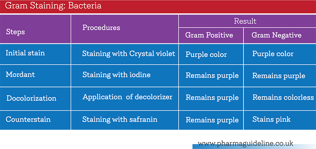
Gram stain is the most useful staining technique engaged in bacteriology, is a differential stain. By using this technique, it is possible to divide bacteria into two different groups- Gram Positive & Gram Negative.

Endospore Staining

Spore stain is a most useful staining technique engaged in bacteriology is a structural stains. By this technique, it is possible to detect the presence & position of endospore in the bacteria.

Responsibilities:
Executive/ Sr. Executive, Microbiology

Initiative of subculture of Microorganisms, perform staining & report preparation.

Manager, Microbiology

Confirm staining techniques, safety report checking, document preservation & application of sound technical information.

Head of Quality Assurance

Approval of SOP

Procedure:

Instructions
  • Standard microorganisms are usually non-pathogenic but those are unscrupulous pathogen. So before handling it, wear protective items such as sterile wear sterile latex free gloves, laboratory coat & eye protection (if necessary).
  • Don’t move vigorously into test area. Move always softly.
  • After completion of subculture or transfer of pellets, disinfect outer surface of the vial or test or plate with 70% IPA.
  • Remove all used items those are directly contacted with standard micro-organisms. Leave it at designated containers safely.
  • Ensure that the waste container is tightly capped until perform autoclaving.
  • Don’t touch the apparatus directly in open hands those are used.
  • Make sure that all personal ornaments, cell phone are left to prevent unauthorized contamination, before entrance into the test room.
Simple Staining
Staining Reagents
Methylene blue Stock Solution :
  • Methylene Blue         2.g
  • Distilled Water          100 ml
Methylene blue Staining Solution :
  • 0.2 % of Methylene Blue Solution  12.5 ml
  • Distilled Water                                87.5 ml
Crystal Violet Solution
  • Crystal Violet            2.0 g
  • Ethyl alcohol 95%    20 ml
  • NH4 Oxalate            0.8 g
  • Distilled Water         100 ml
Carbol Fuchsin(Zeihl-Neelsen)  Solution
  • Fuschin                     1 g
  • Ethanol                     10 ml
  • Phenol                       5 g
  • DistilledWater           200ml

Dissolve Fuschin in alcohol and dissolve phenol in water, then mix the two solutions.

 Apparatus
  • Glass slide 3 x 1 inch
  • Pipettes
  • Gas Burner or spirit Lamp
Staining Technique:
  • Clean & dry microscope slides thoroughly.
  • Flame the surface in which the smear is to be spread.
  • Flame the inoculating loop properly.
  • Transfer a loop full of tap Water to the flamed slide surface.
  • Reflame the loop making sure that the entire length of the wire that will enter the tube has been heated to redness.
  • Remove the tube cap with the fingers of the hand holding the loop.
  • Flame the tube mouth.
  • Touch inoculating loop to the inside of the tube to make sure it is not so hot that it will distort the bacterial cells;  then pick up a pinhead size sample of the bacterial growth without digging into the agar.
  • Reflame tube mouth, replace can, and put tube back in the holder.
  • Disperse bacteria on the loop in the drop of water on the slide and spread the drop over an area the size of a dime.  It should be a thin, even smear.
  • Reflame the inoculating loop to redness including the entire length that entered the tube.
  • Allow the smear to dry thoroughly.
  • Heat-fix the smear cautiously by passing the underside of the slide through the burner flame two or  three times.
  • Test the temperature of slide after each pass against back of the hand.  It has been heated adequately when it feels hot but can still be held against the skin for several seconds.  Excessive heat will distort the cells.
  • Stain smear by flooding it with one of the staining solutions & allowing it to remain covered with stain for time designated below.
  • Methylene blue: 1 minute
  • Crystal violet: 30 seconds
  • Carbol Fuchsin: 20 seconds
  • During staining the slide may be placed on the rack or held in the fingers.
  • At the end of the designated time rinse off the excess stain with gently running tap water.  Rinse thoroughly.
  • Wipe the back of the slide and blot the stained surface with bibulous paper or with a paper towel.
  • Place the stained smear on the microscope stage smear side up and focus the smear using the 10x objective.
  • Choose an area of the smear in which the cells are well spread in a monolayer.  Center the area to be studied.
  • Apply oil directly to the smear, and focus the smear under oil with the 100X objective.
  • Draw the cells observed.


Gram Staining Method
Stain and Reagents
Crystal Violet Solution
  • Crystal Violet                 2.0 g
  • Ethyl alcohol 95%         20 ml
  • NH4 Oxalate                 0.8 g
  • Distilled Water              100 ml
Gram’s Iodine
  • Iodine Crystals              1.0 g
  • Potassium Iodide          2.0 g
  • Distilled Water              100 ml
Docolorizer :
  • Acetone                         50 ml
  • Ethanol 96%                  50 ml
Counterstain :
  • Safranin                         2.5 g
  • Ethanol 95%                 100 ml
  • Distilled Water              100 ml
Apparatus :
  • Glass slide 3 x 1 inch
  • Pipettes
  • Gas Burner or spirit Lamp
Staining Technique
  • Use always young culture of (18 to 24) hours that the differentiation in cell wall structures is retained.
  • Do not use old cultures due to lose Gram Positiveness.
  • Make smear, and dry in air and fix by flaming.
  • Stain with crystal violet for about 30 seconds.
  • Rinse with the water.
  • Cover smear with Gram’s iodine for about 30 seconds.
  • Rinse with the water.
  • Decolorize with 95% ethanol. For a thin smear, 10-20 seconds is long enough; after the proper time interval, alcohol
  • Drippings from the slide are no longer colored.
  • Rinse with the water.
  • Counterstain with safranin solution for 20-30 seconds.
  • Rinse with water and blot dry.
  • Examine under the oil-immersion objectives.
Interpretation of Result :
  • Gram positive bacteria retain the crystal violet dye after de-colorization and appear deep blue or purple colour.
  • Gram negative bacteria are not capable of retaining the crystal violet dye after de-colorization and are counterstained red or pink by the safranin dye.
Endospore staining :
Stain and reagents :
  • Malachine Green solution
  • Malachite green        5 g
  • Distilled Water          100 ml
Counterstain Solution
  • Safranin                   2.5 g
  • Ethanol 95%           100 ml
  • Distilled water         10 ml
Apparatus
  • Glass slide 3 x 1 inch
  • Pipettes
  • Gas burner
Slide Cleaning :
  • Clean the slide by immersion in concentrated Sulphuric Acid saturated with Potassium dichromate for several days for removal of grease from slide.
  • Place drop water on the surface of the slide, if water is spread over its surface indicates the slide is cleaned properly.
Staining Technique
  • Prepare smear, dry in air and fix by flaming.
  • Place the slides on a staining rack.
  • Cover the smear and keep saturated with malachite green 5% aqueous solution and continue heating for 5 minutes.
  • Wash gently with the water.
  • Counterstain with safranin for 30 seconds.
  • Wash with water and blot dry.
  • Examine under the oil-immersion objectives.
Interpretation of result :

The endospore stains green and the remainder of the cell (or a cell without an endospore) stains light red.

The phase microscope is effective in observing the endospore without staining where it appears as a dense white structure in the cell. If a phase microscope is available, observe the unstained endospore in cultures of Bacillus and Clostridium species.

Staining of Microorganism and its Standard Operation Procedure Read More »

Effluent Water Test and Its Standard Operating Procedure

Effluent Water Test ; Purpose:

To ensure that effluent water is analyzed to meet In-house specification in order to minimize environmental pollution.

Scope:

This SOP applies for analysis of effluent water in Quality Control Section and Microbiology Laboratory at XX Pharmaceuticals Limited.

Definition/Abbreviation:  

BOD:  Biological Oxygen Demand

Responsibilities:

The roles and responsibility is as follows:

Microbiologist/Officer, QC

Sample collection and analysis of effluent water & test report preparation.

Manager, Microbiology/Asst. Manager, QC

Ensure sampling, analysis, documentation & application of sound technical information.

Manager, Quality Assurance

Approval of SOP.

Procedure:

Instructions
  • Care should be taken during sampling because effluent water is hazardous and toxic.
  • Wear gloves, mask during sampling and testing.
  • Discard all used materials, glassware, gloves at the end of work after autoclaving.
Sample Collection:
  • Take clean & dry of 2 Liters flask for chemical test sample.
  • Sterilize sampling container at 1210C for 15 minutes for microbiology test.
  • Take effluent water sample at least 2 Liters for chemical test.
  • For microbiology test, take sample into three of 250 ml BOD bottle.
  • Drop cap on the mouth of BOD bottle tightly.
  • Ensure the BOD bottle is free from any bubble.
  • Don’t expose container outside area of Laboratory.
  • Carry out sample into Quality Control & Microbiology Laboratory.
Sample Preservation

Preserve sample at 2 to 80C for not more than 24 hours.

Test Frequency

Carry out test once in every three months.

 Chemical & Microbiological tests as follows

Perform the test as per Analytical Method of Effluent Water.

Report preparation:

Report of the test result in Effluent Water Test Report, Annexure-I.

 Distribution of Result
  • After completion of analysis, inform status to the Engineering Department.
  • If any test result found out of specification, repeat test to be done.
  • If repeat test result found out of specification then inform to Engineering Department for corrective action.
  • After corrective action, collect the sample & carry out the test.

Annexure:

Download the Annexure Here: Report

Effluent Water Test and Its Standard Operating Procedure Read More »

Bioassay Procedure and Its Sop

Bioassay; Purpose

Bioassay; To determine the potency of antibiotics of raw materials & products which are specified in different Pharmacopeia.

Bioassay; Scope

This SOP is applicable for Biological Assay of Antibiotics in Microbiology Section at XX Pharmaceuticals Limited.

Definitions
Microbial Assay

In Microbial assay the potency or concentration of a chemical substance (especially antibiotics) may be determined by its effect on the growth of a defined microorganism.

Responsibilities
Executive/Senior Executive, Microbiology

Assay plate preparation, assay dilution, inoculation, zone reading and report preparation.

Manager, Microbiology

Ensure that all activities of Biological assay, document preservation & application of sound technical information.
Checking SOP that the relevant technical information is applied.

 Bioassay Procedure
Instructions
  • Standard microorganisms are usually non-pathogenic but those are unscrupulous pathogen. So before handling it, wear protective items such as sterile wear sterile latex free gloves, laboratory coat & eye protection (if required).
  • Advised to move softly at Test area not vigorously.
  • After completion of sub-culture or transfer of pellets, disinfect the outer surface of vial or test or plate with 70% IPA.
  • Remove all used items those are directly contacted with standard micro-organisms. Leave it at designated containers safely.
  • Ensure waste container is properly capped until autoclaving.
  • Don’t touch apparatus directly in open hands those are used.
  • To lessen the contamination, make sure that all personal ornaments, cell phone are left before enter into the test room/area’.
Sterilization of Apparatus & Glassware:
  • Sterilize all glassware including Latin Square Plate and others heat stable apparatus at 2000C for 60 minutes in hot air oven using a validated process.
  • Use the glassware when the temperature reduce below 400′
Preparation of Culture Media:
  • Select the media & diluents as per instruction of BP or USP.
  • Prepare 300 ml of particular media as per indication by manufacturer instructions.
  • Bring to boil for dissolve completely.
  • Sterilize at 1210C for at least 15 minutes.
  • Cool media approximately to 450C to 500C
  • Allow to solidify the agar media to prepare the slope.
Preparation of Test solution
  • Prepare different concentrations of Test Solution to determine of the lowest dose for detectable zone of inhibitions.
  • Select & prepare two concentrations of the Test solution as “High Dose” and “Low Dose”.
Preparation of Standard solution
  • Prepare different concentrations of Standard Solution to determine of the lowest dose for detectable zone of inhibitions.
  • Select & prepare two concentrations of Standard solution as “High Dose” & “Low Dose”.
Preparation of Microorganisms Suspension:
For Spore suspension preparation
  • Prepare 140ml of Nutrient agar medium.
  • Aseptically Pour the medium on a petri plate (190mm).
  • Allow the medium to solidify.
  • Keep plate in refrigerator for 30 minutes.
  • After 30 minutes take out plate & streak whole plate with the desired organism aseptically.
  • Keep plate for incubation at 350C for 7 days.
  • After incubation period take out the culture with the aid of sterilized glass beads & pre-sterilized saline water by rotating plate.
  • Pour culture suspension along with few of those glass beads in a 100ml flask containing 50ml of sterilized saline.
  • Heat culture suspension at 700C for 30 minutes in a water bath for spore formation.
  • Cool suspension & then keep inside a refrigerator between (2 to 8)0
  • Don’t use the spore suspension more than 60 days.
For Maintenance of Sub-culture of Vegetative bacteria
  • Before using a culture maintained in a slant, subculture the organism in another slant containing a specified medium.
  • Keep slant for incubation at (30 to 35)0C for 24 to 30 hours.
  • Store the slant in the refrigerator at not more than 7 days.
 Method: Plate Diffusion:
  • Select Latin Square Plate of 12” X 12” size.
  • Place plate on a leveled surface after sterilization.
  • To the medium( 45 to 50)0C add the organism mentioned in above chart for a particular antibiotic in required amount.
  • Shake flask gently to distribute organism throughout the medium.
  • Pour medium on plate & allow it to stand for 30 minutes before placing lid in position.
  • Transfer plate into refrigerator.
  • When required for use, cut cups in agar by means of a sterile cork borer of 8mm diameter.
  • Remove each disc of agar with a “spear” so that the surrounding is not lifted.
Application of solution to Assay Plate :
  • Enter details of the sample numbers, weight & dilutions on the assay report form, after diluting the standard and test solutions, There is an assay report form for each plate.
  • Concentrated solutions are coded with H (High dose) and the lower concentrated solutions are coded with L (Low dose).
  • Apply using a standard (100 ±1) µL the solutions to the assay plate in the order of the design.
  • Starting at top left hand corner & working from left to right across the rows down to the bottom right hand corner.
  • Once started, plating out should be continuous, as it is important that solutions are placed in cups at regular interval.
  • Keep the plate about one hour for proper diffusion.
  • After diffusion lid the plate with glass lid.
Incubation of Assay Plate

Incubate assay plate for (16 to 20) hours at 370C for antibiotic or 250C for antifungal.

Measurement of the Zone Diameters
  • Place the assay plate on a photographic light box.
  • Measure zone of inhibition using Varnier calipers.
  • Start at top left hand corner and measure the diameter of the zone accurately.
  • Continue measuring zones from left to right on row 1, then right to left on row 2.
  • Repeat the procedure until all 64 zones have been properly measured.
Bioassay; Calculation of Potencies:
  • Sum high & low doses for each standard ( S1&S2)
  • Sum high & low doses for each test sample (T1& T2)
  • Substrate test treatment totals from standard treatment total. This will give a plus or a

Minus figure = D, D = (T1 +T2)-(S1 +S2).

  • B = (Sum of high doses of test, T1 – Sum of low doses of test solution, T2) + (Sum of high doses of

Standard, S1– Sum of low doses of standard, S2)

  • Calculate the “Dilution factor of high-test sample (F)” by dividing “Weight of sample” taken by “Total

Volume of dilution” up to high doses.

  • Log ratio of dilution (I) = Log (High dose concentration ÷ Low dose concentration)
Actual weight

Calculate “Potency of High Standard (H) = ———————– X. High dose concentration                                                             Theoretical weight

Potency (P) = Antilog (D/B x I) x F x H.

Report the result in Biological Assay Report, Annexure-I.

Download Annexure: Bioassay Report

Bioassay Procedure and Its Sop Read More »

Environmental Monitoring Procedure and its Sop

Environmental Monitoring; Purpose

Environmental Monitoring; To describe the procedure for Environmental monitoring.

Scope

Environmental monitoring in  XX Pharmaceuticals Ltd

Definitions
Environmental monitoring:

Monitoring of viable and non-viable quality of a controlled environment

Particulate count:

[] Enumeration of non-viable particulate of specific size from a particular volume of air of a controlled
Environment.
[] Settle Plate: Exposure of petri-plates of nutrient media in a controlled environment to estimate viable
Microorganisms from the environment.
[] TSA: Tryptone Soya Agar
[] CFU: Colony Forming Unit

Responsibilities

The roles and responsibility is as follows:

Lab. Attendant

Room preparation for Test

Executive, Microbiology

Carry out the test and incubation & documentation

Sr. Executive, Microbiology

To ensure test, incubation, report checking, document preservation & application of sound technical information.

Head of Plant

Review of the SOP & the relevant technical information is applied.

Head of Quality Assurance

Take initiative to Approve of this SOP.

Procedure:

Instructions
  • Don’t rub during contact plate sampling.
  • Use the sterile filter holder.
  • Place the filter paper on the filter holder carefully under laminar airflow.
  • Try to avoid unwanted personal contamination during air sampling.
  • Monitoring to be carried out before production hours. (Sterile products)
Non-viable Particle monitoring:
  • Bring the laser particle counter into the specific monitoring area.
  • Enter into clean room, wearing approved designated dress. When entering into the clean area, before taking reading make sure that all the doors remain closed.
  • Operate the Particle counter following approved SOP.
  • Take count of different several position for each room as indicated in sampling point. Count the number particles (5µ and 0.5 µ) form each sampling point. The average of the counted particle indicates the total particles of a room.

The minimum sampling time should be as per following formula (Following EN ISO 14644-1)

Vs=(20/Cn,m ) x 100

Where,

  • Vs = is the minimum single sampling volume per sampling point, expressed in liter.
  • Cn,m = is the class limit (number of particles per cubic meter) for the largest considered
    particle size, specified for the relevant class.
  • 20 = is the defined number of particles that could be counted if the particle concentration
    were at the class limit.
Specification: Follow Annexure-III.
Write down the result in the format of Annexure-I.


Environmental Monitoring

Viable count:

Settle Plate
  • Prepare, sterilize & dispense the media TSA into the petriplates and pre-incubate the plates.
  • Check the pre-incubated plates for any evidence of microbiological contamination under the LAF bench.
  • Discard the plates contains microbiological growth.
  • Decontaminate the external surface of petriplates with sterile cloth /cotton soaked in a sanitizing agent (70% IPA).
  • Place required number of petriplate in a sterilized /sanitized SS container; close the lid of SS container.
  • Transfer SS box to the area to be monitored.
  • Enter respective area as per the SOP of Entry and Exit procedure.

Mark Petri plates with the following details-

  1. Name of the sampling point
  2. Room No.
  3. Date of exposure
  • Place petriplates on corresponding designated plate exposure area and remove
    the upper lid of the petriplates. Note the beginning time of the exposure and write it on
    the plate.
  • Place upper lid on edge of petriplates in slanting position.
  • Expose media plates at sampling point for Maximum 4 hours.
  • After completion of exposure, close petriplates with lid.
  • Collect petriplates in the SS container and bring back exposed plates to microbiology Laboratory.
  • Incubate all exposed petri plates in inverted position in laboratory Incubator.
  • Incubate at (22.5 ± 2.5)°C for mold and Yeast for 72 hours followed by another 48 hours for bacteria at (32.5 ± 2.5)°C. After incubation count the number of colony forming units (CFU) per plate per sampling point with colony counter.
  • Incubate an unexposed media filled petriplates along with the exposed plates and mark it as negative control.
  • Count the number of Colony Forming Units (CFU) per plate per location with colony counter. The average of colony indicates total CFU of a room.
Write down observation in the format attached as Annexure-II.

Air Sampling:

  • Set instrument for desired sampling time & flow rate as per Approved SOP.
  • Take air sampler to location where air is to be sampled, hold it with filter facing at required direction and take air sample following approved SOP.
  • Collect filter under laminar airflow & place it inside on petriplates containing TSA[Tryptone Soya Agar ] media.
  • Incubate all exposed petri plates in inverted position in the microbiology laboratory Incubator. Incubate at the (22.5 ± 2.5)°C for mold & Yeast for 72 hours followed by another 48 hours for bacteria at (32.5 ± 2.5)°C. After incubation count the number of colony forming units (CFU) per plate per location with colony counter.
  • Follow sampling location.
  • Record observation in format attached as Annexure-II.
  • Calculate number of organisms per cubic meter of air. Average colony indicates total cfu of a room.

Surface monitoring (By swab sampling):

  • Use sterilized swab or sterilize the swab in the autoclave.
  • Place required number of sterilized swab sticks with tube containing 5 ml of sterile saline
    solution in a Sterilized SS container & tightly secure lid of the SS container.
  • Transfer SS box to area to be monitored.
  • Enter respective area as per SOP of entry and exit procedure.

Remove swab sticks with tubes from the SS box & take it to the location to be monitored and Mark tube with following details:

  1. Location number or Name of the location
  2. Swab No.
  3. Date of sampling
  • Hold swab stick from bottom & place tip on surface to be monitored. Gently wipe the swab bi-directionally to cover an area about 25 cm2.
  • Perform swab sampling for each defined locations.
  • Keep swab sample standing in the SS container & bring it to Laboratory.
  • Gently vortexes swab sampling tube containing swab stick & pour contents in filter holder funnel and filter it. Uses the respective SOP.
  • Rinse tube with 3 x 10 ml of sterile saline solution.
  • Filter test sample under partial vacuum.
  • Rinse Funnel with the portions of sterile Purified Water. This flushes residue from walls of funnel & helps to secure a uniform distribution of colonies on filter surface. Upon completion of rinse & filtration process, shut off vacuum.
  • Transfer membrane filter with sterile smooth-tip forceps on to Microbial content test agar (TSA) plate.
  • Place filter with a rolling motion to avoid entrapment of air.
  • Keep a negative control by filtering 100 ml Purified Water(Sterile) before filtering actual test sample.
  • Pass 20 to 30 ml of sterile Purified Water through funnel, between different test samples when the same funnel used for multiple sample.
  • Incubate all exposed petriplates in inverted position in microbiology laboratory Incubator. Incubate at (22.5 ± 2.5)°C for mold and Yeast for 72 hours followed by another 48 hours for bacteria at (32.5 ± 2.5°)C. After incubation count number of colony forming units (CFU) per plate per location with colony counter.
  • Count number of colony forming units (CFU) per plate per location with colony counter.
Note down the observation in the format attached as Annexure-II
Surface monitoring (By contact plate):
  • Fill Petridishes with TSA culture medium & pre-incubate.
  • Take off the lid of plates.
  • Invert & press agar surface for 10 seconds onto surface to be examined.
  • Replace lid & mark plate with appropriate data.
  • Clean sampling area on surface in order to remove any remaining of agar.
  • Return plates to laboratory.
  • Incubate all exposed petriplates in inverted position in microbiology laboratory Incubator. Incubate at (22.5 ± 2.5)°C for mold and Yeast for 72 hours followed by another 48 hours for bacteria at (32.5 ± 2.5°)C. After incubation count number of colony forming units (CFU) per plate per location with colony counter.
  • Take count of visible colonies within mean of plate
  • Express results as CFU per contact plate.
  • Perform appropriate identification of germs found by surface monitoring..Classified area should free of pathogenic microorganism.
Note down the observation in the format attached as Annexure-II.
Sampling Location:

Determine Number of sampling location by following formula.

NL    = √A

Where,

NL   = the minimum number of sampling locations, rounded up to a hole number.

A= Area of the clean room or clean air controlled space in m2.

Environmental Monitoring Procedure as per Pharmacopeia

Download All Annexure Here: Environmental Monitoring

Schedule: Environmental Monitoring

Environmental Monitoring Procedure and its Sop Read More »

Suitability of Microbial Count Method & its SOP

Suitability of Microbial Count Method; Purpose

Suitability of Microbial Count Method; To confirm the ability & the suitability of the test to detect microorganisms in the presence of a product or Raw Materials as per the In-house or Pharmacopoeia specifications.

 Scope

This SOP is applicable for Microbial Count Method Validation in Microbiology Section of XX Pharmaceuticals Limited.

 Definitions

Microbial count Suitability: Microbial Count suitability is to confirm that the test dilution is free from any type of interfering substances or antimicrobial properties that will recover by dilution or the addition of a neutralizer to detect microorganisms in the presence of the product.

  • CSDA: Casein Soybean Digest Agar
  • CSDB: Casein Soyabean Digest Broth
  • LAF: Laminar Air Flow
  • SDA: Sabouraud Dextrose Agar
  • SDB: Sabouraud Dextrose broth
Responsibilities

The roles and responsibility is defined as follows:

Executive/ Sr. Executive, Microbiology

Preparation & inoculation of standard culture, carry out test & arrange appropriate document preparation.

Asst. Manager/Manager, Microbiology

Ensure of method suitability, documentation and application of sound technical information.

Head of Quality Assurance

Taking Initiative to Approval of this SOP

Procedure

Instructions
Safety Precautions
  • When enter into the test area, wear sterile latex free gloves, Laboratory coat and eye protection (if required).
  • To prevent unauthorized contamination, make sure that all personal ornaments, cell phone are left before enter into the test room. The use of Cellular phone in the test room is strictly prohibited.
  • Don’t move vigorously into the test area. Move always gently.

General Requirements

Glass Apparatus
  • Pipette 2 ml, 10 ml
  • Sterilized 90 mm Glass Petridish
  • Screw capped Conical Flask 100 ml
  • Screw Capped Test Tube
  • Volumetric Flask 500 ml
  • Volumetric Flask 1000 ml
Media and Reagents
  • Casein Soyabean Digest Agar(CSDA)
  • Casein Soyabean Digest Broth(CSDB)
  • Meat peptone
  • Neutralized Peptone
  • Sabouraud Dextrose Agar
  • Sabouraud Dextrose Broth

These media can be purchased from commercially available manufacturers.

 Others Requirements
  • 45 µm Membrane Filter
  • 70% IPA or ethanol
  • Filtration Unit( sterilized filter disk and filtering funnel)
  • Forceps
  • Glass spreader
  • Scissor
  • Surgical Cotton
  • Surgical Gloves
General Procedures
Test Conditions
  • Wear gloves, mask/beard mask Headgear before entrance into Testing Room.
  • Disinfectant hands, the outer surface of test sample, LAF workstation with the help of 70% IPA or ethanol before starting the test.
  • Carry out the test under LAF to avoid any type of contamination.
  • Monitor the test area microbiologically using Microbial Air Sampler at each working day.
Culture Media Preparation
  • Prepare the different culture media as per the requirement.
  • Weigh the mentioned amount as per manufacturer label into appropriate flask.
  • Bring to boil completely to dissolve the media.
  • Sterilize at 1210C for 15 minutes or as per Manufacturer label.
  • Store the prepared culture media in air tight flask at controlled environment.
  • Store prepared agar media at (2-8)0C
  • Preserve dehydrated culture media up to its expiry date.
  • Never use expired culture media.
  • Use the agar media when the temperature reduce to 450C and cools in case of the broth media.
Stock Buffer Solution
  • Take 34 g of Potassium Dihydrogen Phosphate[KH2PO4] in a 1000 ml volumetric flask.
  • Dissolve in 500 ml Purified Water, adjust to pH [7.2 ± 0.2] then dilute to 1000 ml with Purified Water.
  • Dispense 90 ml into each screw capped flask[Approx. 11 containers].
  • Sterilize at 1210C for 15 minutes.
  • Store the prepared buffer at 2-80C for a validated period.
Glassware Cleaning & Sterilization
  • Clean glassware by 1% detergent initially then rinse with sufficient tap water.
  • Rinse finally with sufficient Purified Water to remove the residual content of detergent.
  • Sterilize glassware at 2000C for 1 hour.
 Sample Preparation
Water-soluble samples

Take 1 gm or 1 ml into the 9 ml CSDB [Casein Soybean Digest Broth] or phosphate buffer or Sodium chloride peptone solutions.

Water-insoluble samples

Take 1 g or 1 ml into the 9 ml CSDB [Casein Soybean Digest Broth] or phosphate buffer or Sodium chloride peptone solutions and add 0.1% Polysorbate 80.

Fatty Products

Dissolve with Isopropyl Myristate sterilized by filtration with low amount of Polysorbate 80 or anther non-inhibitory sterile surface active agent  or necessary heat not more than 450C.

Inoculation and Dilution:
  • To maintain of not more than100 cfu, add adequate volume of suspension of inoculums to the sample.
  • Add the inoculums suspension not more than 1% of diluted product.
  • Prepare lowest possible dilution for acceptable microbial recovery of sample.
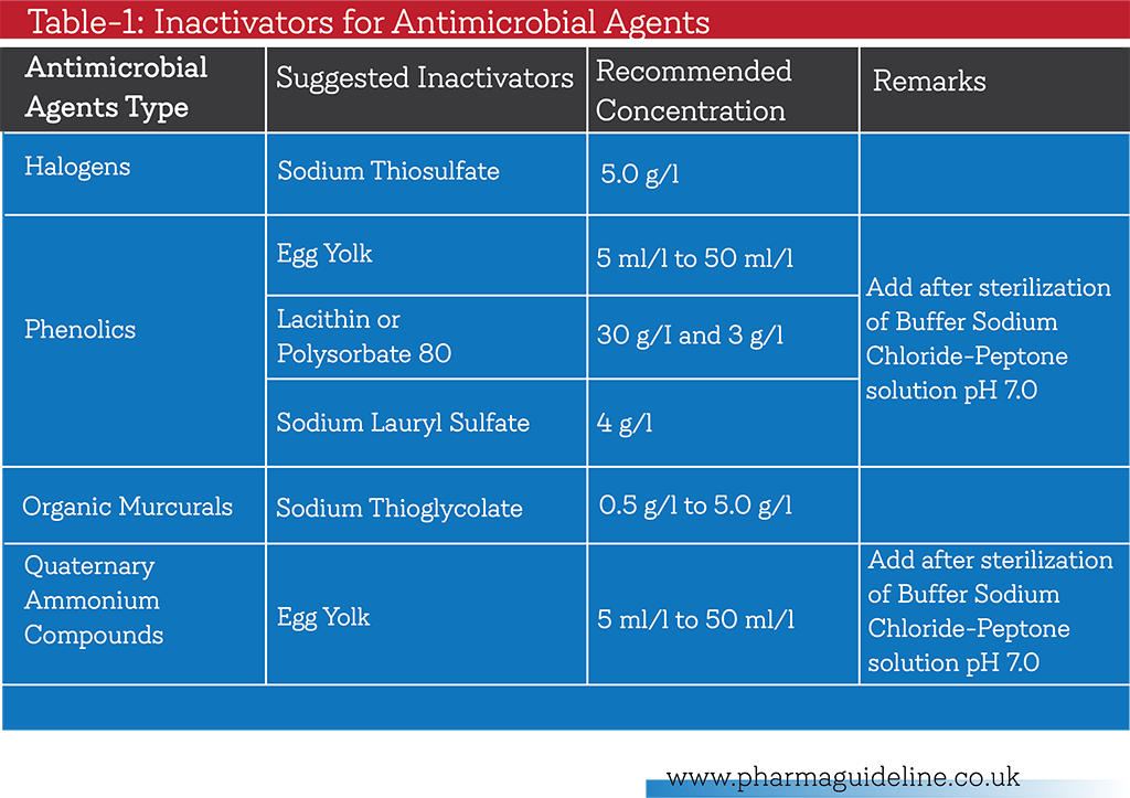
  • Add neutralizer for removal of Interfering factor when [If] the sample contains any antimicrobial properties.

Follow Table-1 for the inactivators of  Antimicrobial agents.

If growth is inhibited, then increase use of diluents or membrane filtration or combination of all above.
 

Suitability of Microbial Count Method


Suitability of Counting Method:

[] Membrane Filtration Method

[] Most Probable Number Method

[] Pour Plate Method

[] Surface-spread plate Method

Pour Plate Method :
  • Add 1 ml prepared sample to the 90 mm diameter Petridish
  • Pour (20~25) ml of Sabouraud Dextrose Agar(SDA) for fungi and Casein Soybean Digest Agar(CSDA) for Bacterial count both media being not more than 450
  • If larger Petridish [more than 90 mm diameter] is used amount of media to be increased accordingly.
  • Perform plate count methods at least in duplicate for each medium and use the mean count of the result.
  • Inoculate the microorganisms not more than 100 cfu as indicated Table 2.
  • Mix properly & incubate CSDA plate at (30~35)0C for 3 days, SDA plate at (20~25)0C for 5 days.
  • After incubation, count the colony on each plate.
  • Take arithmetic mean of the count per medium.
Surface-Spread Plate Method
  • Add (20~25) ml of SCDA and SDA to 90 mm diameter petridish at least duplicate.
  • Allow to solidify and dry the plate under Laminar Air Flow cabinet or incubator.
  • Spread not less than 0.1 ml (equal to or less than 100 cfu) of each microorganisms as indicated in Table 1 on the surface of medium
  • Incubate at CSDA plate at (30~35)0C for 3 days, SDA plate at (20-25)0C for 5 days.
  • After incubation, count the colony of each plate.
  • Take arithmetic mean of the count per medium.
Membrane Filtration Method
  • Use the membrane filter which nominal pore size is not more than 0.45µm.
  • Filter prepared sample through membrane.
  • Rinse membrane filter with sterile 0.1% meat peptone solution or any others suitable diluents for neutralization of the sample.
  • Add inoculums to filter as indicated in Table 1 & rinse again.
  • Transfer filter on the surface of CSDA plate & SDA plate.
  • Incubate CSDA plate at (30 to 35)0C for 3 days, SDA plate at (20 to 25)0C for 5 days.
  • After incubation, ten count the colony of each plate.
  • Take arithmetic mean of the count per medium.
Test Control

Negative Control: Use diluents in place of test preparations. There must be no growth found in the negative control. If found any growth in the negative control, then the test is invalid & repeat the test.
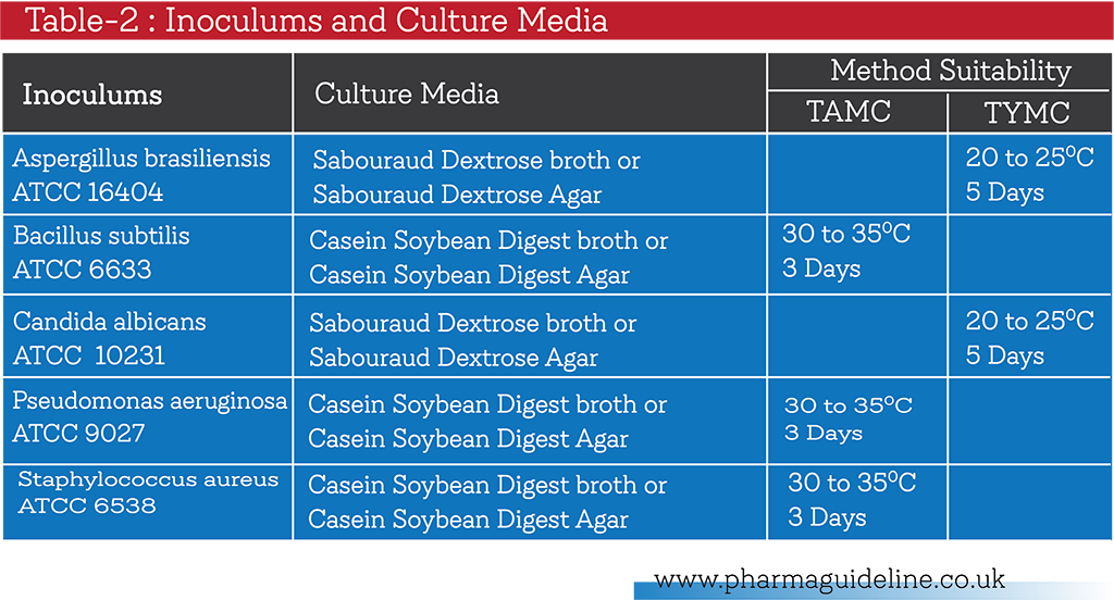
 

Suitability of Microbial Count Method

Result and Interpretation

Mean count of any of test microorganisms not differing by a factor greater than 2 from value of the control defined in absence of product must be obtained.

Repeat test by increasing neutralizer or dilution or any treatment for overcome of antimicrobial properties of products. If the above criteria cannot be met for one or more microorganisms.

Test Report Preparation

Prepare Report the result in Suitability Report of Microbial Count Method, Annexure-I.

Annexure

Annexure-I: Suitability of Microbial Count:
Suitability Test Report of Microbial Count Method

Suitability of Microbial Count Method & its SOP Read More »